How does MRI work ?
Magnetic Resonance Imaging (MRI) also called as Nuclear Magnetic Resonance Imaging (NMRI) or Magnetic Resonance Tomography (MRT) is an imaging technique which uses magnetic field to generate images of the body. MRI can be easily conceptualised once the Physics and Chemistry behind it is understood. So let’s begin !
MRI – Physics
We all have read a chapter called Magnetism in our +2s. Our body is made up of elements which are ultimately made up of atoms. Now an atom has a nucleus containing positively charged proton and neutron with no charge. The nucleus is surrounded by negatively charged electrons. The positive charges are continuously moving inside the nucleus i.e. they have an angular momentum. This angular motion is called spin of the particle (electron, proton or neutron)
As a dictum we know that “every moving charge creates a magnetic field.”
Hydrogen Atom : The human body is composed of 70% water. MRI relies on the properties of H atom to produce images. Single proton in H atom produces a magnetic field (called as magnetic moment or dipole moment) being a spinning charged particle. Normally the protons are oriented randomly, hence there is no overall magnetic field (They mutually cancel out)

Why electron of H atom is not used for MRI ?
One might wonder that a H atom has two charged particles – electron and a proton, both possess a magnetic field, so why we specifically use magnetic field of proton for MRI.
The magnetic dipole moment is dependent on the mass and charge of an elementary particle. Electron is not affected by magnetic field produced by MRI because the dipole moment of electron is far greater than a proton (X 658 times stronger) due to greater mass of proton (X 1836 times).
This means that normally Electromagnetic waves (radio waves) can penetrate tissues normally to excite H nuclei, but to excite the electronic magnetic moment the energy required is 658 times more becoming this way of microwave in EM spectrum.
But microwaves can penetrate only small centimetres of tissue making it unsuitable for study due to high energy deposit on surface.
Hence due to this reason we don’t use electron of H atom in MRI and rather use proton for the purpose of imaging.
Components of MRI Machine
Now let’s have a look at what is inside a MRI machine.
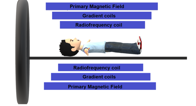
- Primary Magnetic Field : This magnetic field is produced by a superconducting magnet of 1.5 – 3 Tesla strength.
- Gradient Coil : It is used to produce a variation (gradient) in the magnetic field in X , Y and Z axes.
- Radiofrequency (RF) Coils : These are simply loops of wire that transmit the RF signal and also receive the return signal from the body of patient.
Once a primary magnetic field (B0) is applied the H atoms align parallel or antiparallel to the direction of application of magnetic field. This process is called Longitudinal Magnetisation.


It should be noted that a greater proportion aligns parallel (low energy state) than antiparallel (higher energy state) to the primary magnetic field B0.
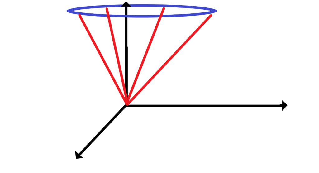
Precession in MRI
Precession :Protons spin around the long axis of the primary magnetic field which is called Precession. It can be further understood as the protons rotate inside the nucleus of H atom (spin) and the whole H atom (and thus the protons inside the atom too) rotate along the direction of magnetic field (precession).
When protons precess together they are said to spin In-phase and to the contrary they are called Out of phase when they precess randomly. Normally at any given time protons precess haphazardly (due to entropy) and become aligned only when an external field is applied upon them. The frequency of In-phase precession when an eternal field is applied on the protons is called Larmor or precessional frequency after the Irish physicist and mathematician Sir Joseph Larmor.

Longitudinal and Transverse Magnetization
Now when the protons are precessing in a particular axis they generate a magnetic field in that corresponding axis. The magnetic field generated by the protons in the Longitudinal axis parallel to axis B0 is called Longitudenal Magnetization and that perpendicular to B0 is called Transverse Magnetization.
Flipping of protons
Now if we switch on the Radiofrequency Coils (RF Coils) for a brief period of time it will apply an external magnetic field which has the same precession frequency as of the protons. This second magnetic field (also called as RF Pulse) will supply some of its own energy to the already precessing protons. This energy is specifically taken up by the protons precessing at low energy states to jump from the lower energy states to higher energy states (Flipping of protons) thus decreasing their Transverse magnetization and increasing their Longitudinal magnetization.
Not only do the protons flip but they also start precessing In-phase under the influence of RF pulse.
Relaxation
In the previous section we saw that the RF pulse provided energy to the protons which helped them to flip to higher energy states and as a result there was a growth in Longitudinal magnetization. Now as the protons in the higher energy state are not stable (they encounter steric hindrances) hence they start losing their gained energy. In this process many protons start falling back to the lower energy state which causes a decrease in Longitudinal magnetization and regrowth of Transverse magnetisation.
This whole process by which protons reach back to their equilibrium state is called Relaxation and the time taken in this process is called Relaxation time.
There are two processes that happen during the whole relaxation process –
- T1 Relaxation
- T2 Relaxation
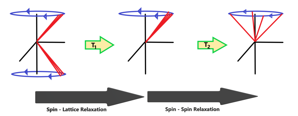
T1 Relaxation
T1 Relaxation happens when protons flip back to their original state by losing some energy. As there is a change in energy states of proton, T1 relaxation is also called as Spin – Lattice Relaxation. The ‘T1’ stands for the ‘Thermal relaxation’.
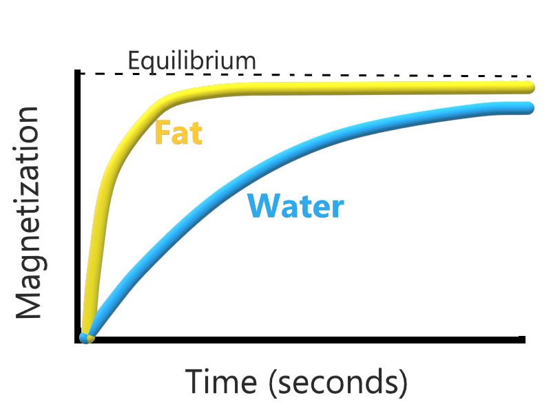
T1 relaxation majorly depends on the composition of the tissue because steric hindrances affect the loss of energy of protons (we’ll understand this in detail in T2 weighted MRI section).
T2 Relaxation
After RF Pulse the protons that were In-phase begin to dephase out of the larmour frequency in the transverse axis. This relaxation is called T2 or Spin – Spin Relaxation.
Just like T1 relaxation, T2 relaxation also varies with the composition of the tissue.
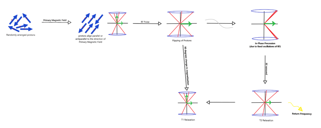
Summary of Processes occurring in MRI