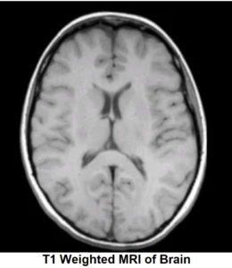
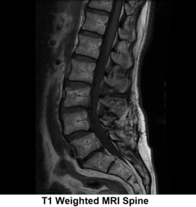
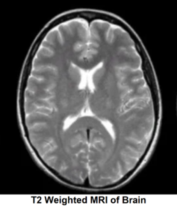
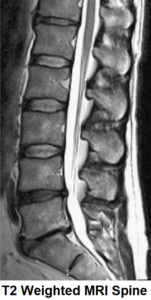
How are T1 and T2 weighted images generated ?
Each one of us has used a camera for capturing photographs and videos. Just like a camera which has multiple settings and adjustments for capturing different versions of an image, an MRI machine also uses a combination of various RF pulses and gradients to generate particular image appearances called as MRI sequences. T1 and T2 weighted images are two of such MRI sequences.
‘Weighted’ term in MRI means that the contrast and brightness of the image (which we see on MRI film) depends on the particular property of tissue. A T1 weighted MRI depends on how fast or slow T1 relaxation happens in the protons of a tissue whereas T2 weighted MRI depends on how fast or slow the T2 relaxation happens in the tissue.
Let’s have a detailed look on how these MRI image sequences are generated.
For better understanding let’s decode T2 weighted MRI first.
T2 Weighted MRI
MRI Spin Echo Effect

T2 Weighted Sequence
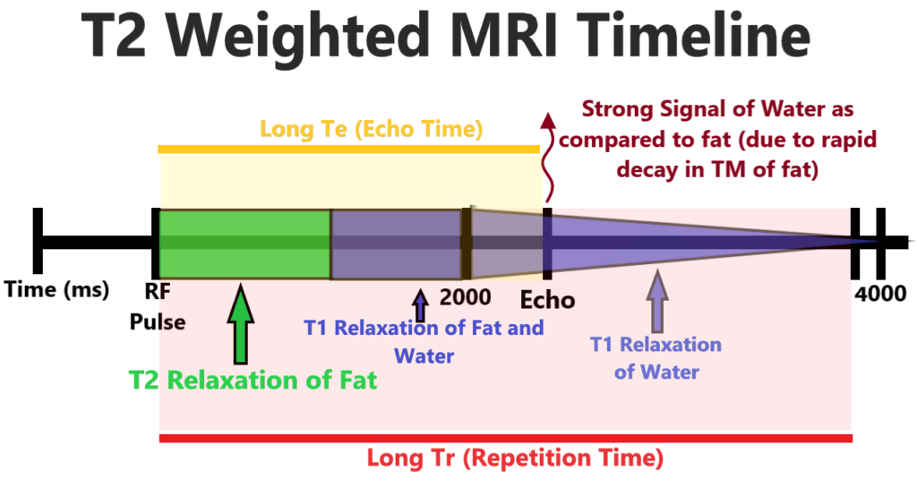
When we put a RF pulse all protons absorb energy, flipping to higher energy state and spin together to produce a 900 pulse or Transverse magnetization(TM). If we wait for a sufficient time, protons will move apart in T2 relaxation and TM will decay. Protons of free fatty acid being relatively fixed in position, decay rapidly as spins push away from one another. They also give up their absorbed energy as they fall back to the baseline in a T1 relaxation, depositing heat energy to the surrounding tissues thus regrowing Longitudinal magnetization(LM).
On the other hand protons in free flowing water can hold energy and continue to spin in phase thus maintaining TM. At this point if we turn on the measuring coil and measure the signal coming out of the relatively large TM in H2O will give a strong signal while smaller/absent TM in fat will give a weak signal. By convention, strong H2O signal is white and weak signal by fat is grey or black in color.
Hence to accentuate different T2 relaxations in of protons in our body we would wait a long period between RF pulses (Long TR) and wait a long time to listen for the return signal or Echo(Long TE).
T1 Weighted MRI
MRI Spin Echo Effect

T1 Weighted Sequence
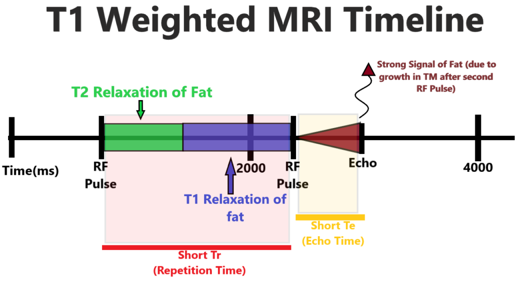
To accentuate T1 relaxation we again put a 90o resonance RF Pulse which flips the protons into high energy state and pushes them in phase to produce TM.T2 relaxation occurs as the protons move apart faster in fat than in water and then fall back into the low energy state dissipating the absorbed energy as heat into the surrounding tissues and thus regrowing LM. Because protons in water are free to move they tend to hold energy and stay in high energy state with little regrowth of LM, whereas tightly packed fat protons more rapidly give up that energy and return to low energy state thus rapidly regrowing LM.
If we quickly put another resonant 90o RF Pulse, the fully recovered fat protons will produce a large TM and a strong measure signal that we can record if we listen to return signal or Echo shortly after second RF pulse. However protons in water are still in a higher energy state with little regrowth of LM.
Therefore the new pulse flips more of low energy protons to high energy state and can only produce a small TM and a net LM oriented 180o or directly opposite the main magnetic field. This will give a low amplitude wave when we listen echo of return signal. The protons in Water are saturated and can no longer produce a strong TM.
Therefore to accentuate the differences in T1 relaxation between protons in fat and water we have to rapidly put in resonating RF Pulse (Short Tr) and quickly listen for return signal( Short Te).
Identification of T1 and T2 Weighted MRI
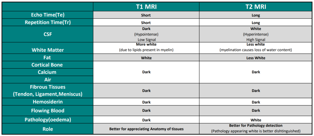
For Identification, at first one should look at the Bone. If the bone is Dark and Scalp fat is white, it’s an MRI image. Observe the Organ (brain, heart, liver, biliary tract, kidneys, spleen, bowel, pancreas, adrenal glands etc.) and Section in which the organ has been represented (axial, saggital, coronal).
After this one should observe the colour of CSF and white matter. Dark appearing CSF and white matter appearing more white indicates that the section is a T1 Weighted MRI and with white CSF and white matter being darker than grey matter, the MRI sequence is T2 weighted.
